AI CERTS
2 hours ago
AI-Enhanced Cardiac Imaging: Inside Ventripoint’s VMS+ 4.0
Moreover, the system promises point-of-care volumetrics for left and right chambers alike. Its FDA clearance signals a broader shift in Cardiac Imaging toward AI-enhanced ultrasound. However, real value emerges only when clinical evidence, workflow gains, and regulatory milestones align. The following report unpacks the technology, data, competition, and remaining questions for enterprise buyers. Transitioning from single-chamber to whole-heart analytics could reshape cardiac service lines significantly. Therefore, understanding VMS+ 4.0 helps leaders plan investment, training, and future imaging strategies.
Echo AI Market Shift
Global ultrasound vendors now invest heavily in automated quantification. Meanwhile, health systems push for faster exams to ease magnetic resonance backlogs. Market research projects double-digit growth for AI echocardiography platforms through 2030. However, few solutions reconstruct the entire heart rather than single chambers. Ventripoint positions its Knowledge-Based Reconstruction workflow as that differentiator. Consequently, executives monitor how whole-heart metrics influence downstream Cardiac Imaging demand.
Independent studies suggest volumetric echo could reduce follow-up resonance referrals by up to 30 percent. Furthermore, payers explore reimbursement models rewarding point-of-care volume measurement. Steady regulatory approvals add confidence to early adopters despite limited longitudinal outcomes. These trends set the stage for whole-heart platforms to capture share. In summary, the market now rewards solutions that cut scan times without sacrificing accuracy. Next, we examine what distinguishes the latest VMS+ release technically.
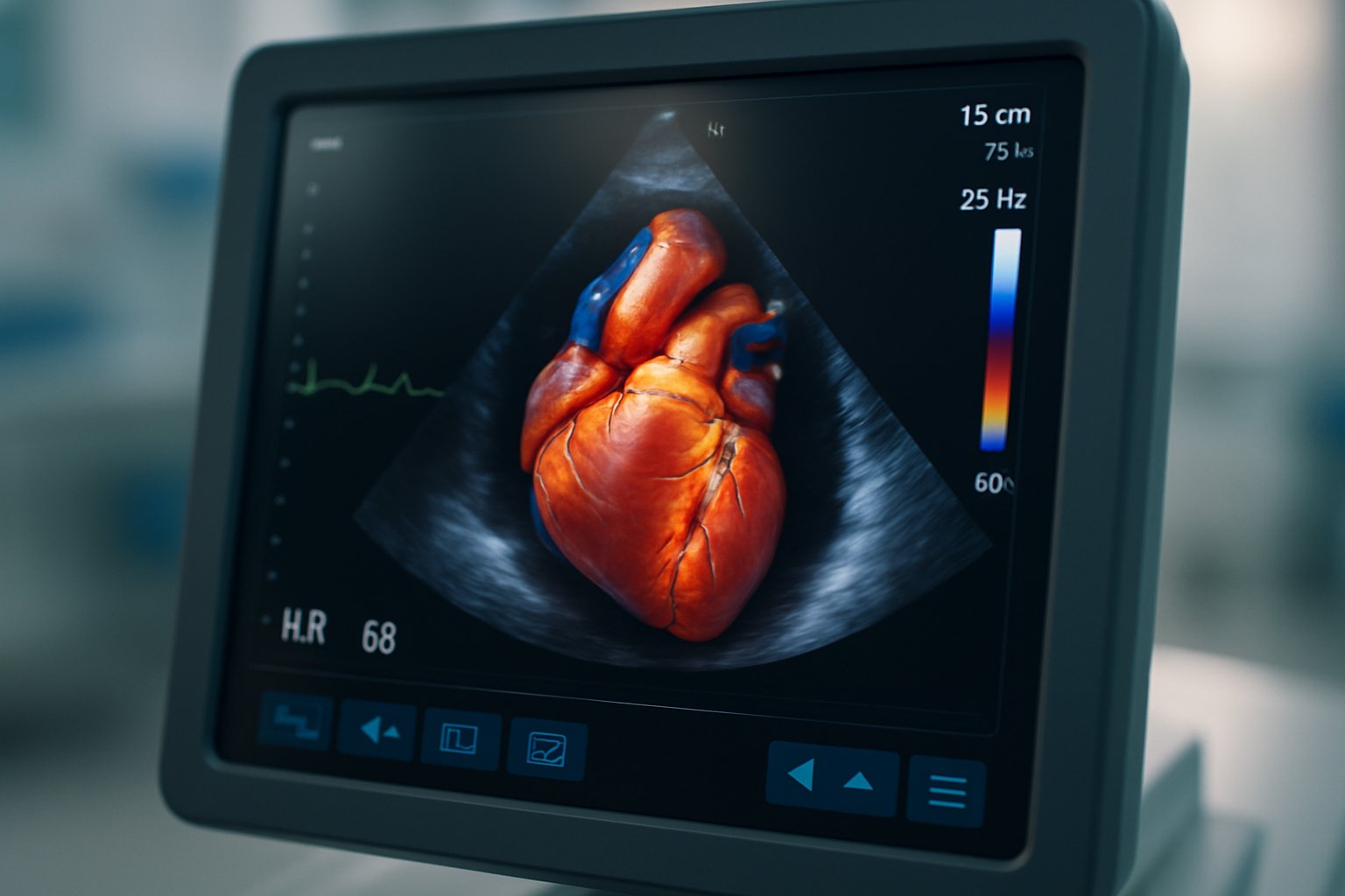
VMS+ 4.0 Overview
VMS+ 4.0 pairs software with small probe-mounted sensors. The sensors record transducer orientation during standard B-mode sweeps. Subsequently, AI algorithms map anatomical landmarks onto a library of MRI-derived shapes. This mapping produces a patient-specific 3D mesh for all four chambers. Automated point placement, introduced in 4.0, accelerates segmentation and reduces training burden. Moreover, magnet-free sensors now allow imaging of pacemaker patients, a previously excluded group. Therefore, hospitals see an opportunity to serve device carriers without costly alternative referrals.
Processing runs on a dedicated workstation but accepts video feeds from most cart manufacturers. Compatibility with existing fleets eases capital budgeting for Cardiac Imaging managers. However, independent confirmation remains advisable before citing institutional endorsements. Key metrics in the FDA summary show volumetric error below five percent. The reference modality was MRI across multiple bench datasets. Such precision supports routine surveillance where small volume changes guide therapy. Thus, version 4.0 advances both usability and eligible patient pool.
- AI-assisted landmark placement for faster segmentation
- Magnet-free probe sensors compatible with pacemakers
- Enhanced reporting with whole-heart volumetric dashboards
Early workflow gains encourage broader evaluation, as the next section shows.
Clinical Evidence Snapshot Today
Peer-reviewed data underpin the system’s claims. A 2021 multicentre paediatric study analysed 545 healthy children using KBR technology. Investigators reported intra-observer ICCs up to 0.998, indicating high reproducibility. Additionally, feasibility exceeded 95 percent across age groups. In contrast, comparative trials with MRI highlight nuanced performance. Volumes correlate strongly, yet ejection fraction agreement varies, especially in complex congenital anatomy.
Nevertheless, authors conclude the tool is suitable for serial follow-up in stable patients. Another JASE study found right-ventricular 3D reconstructions tracked gold-standard volumes within five millilitres. However, systematic biases surfaced near surgical decision thresholds. Consequently, centres continue to confirm borderline findings with conventional Cardiac Imaging. The platform remains complementary rather than a universal replacement. These evidence points frame realistic expectations for clinicians assessing adoption. Next, attention shifts to workflow improvements driving efficiency gains.
Workflow And Sensor Advances
Time pressure in echo labs makes workflow paramount. Version 4.0 shortens data acquisition to under five minutes per patient. Furthermore, AI-assisted point placement cuts manual clicking by 40 percent, according to company testing. Consequently, repeat scans drop, saving sonographer bandwidth. Magnet-free sensors also remove ferromagnetic interference issues present in earlier generations.
Ventripoint highlights easier sterilization because sensors detach quickly. Moreover, the absence of magnets permits use around implanted defibrillators and pacemakers. These innovations align with infection-control and patient-safety initiatives. Importantly, data sets export to common PACS formats, maintaining longitudinal Cardiac Imaging archives. Therefore, IT teams avoid proprietary lock-in while gaining structured volume metrics. With workflow covered, the competitive landscape warrants examination.
Competitive Landscape Update Now
More than 500 AI devices now hold FDA clearance. However, most focus on acquisition guidance or left-ventricular analysis. Philips, GE, and Canon sell consoles with onboard 3D quantification modules. Dedicated software vendors, including TomTec and DiA, offer cloud post-processing. Nevertheless, few platforms recreate every chamber from 2D echoes. VMS stands apart in Cardiac Imaging through its knowledge-based template library originally derived from magnetic resonance scans.
Ultromics, Caption Health, and others target different steps in the workflow. Consequently, hospitals may deploy complementary solutions rather than a single supplier strategy. Licensing models vary, so rigorous cost comparisons remain essential. These dynamics influence procurement but do not override clinical evidence needs. Regulatory milestones now provide another differentiator.
Regulatory And Adoption Milestones
FDA clearance K241222 arrived on February 26, 2025 following rigorous performance reviews. Moreover, CE marking landed in October 2024, enabling European distribution. Canada also issued a medical device licence, extending North American reach. Subsequently, Duke University announced purchase of a second unit early 2024. Seattle Children’s and Mayo Clinic appear in company marketing, though independent confirmation is pending. Nevertheless, multiregional clearance reassures risk committees evaluating deployment.
Hospitals with limited magnetic resonance capacity may benefit most, especially congenital programs. Additionally, pacemaker cohorts finally gain volumetric follow-up without scanning contraindications. Professionals may validate skills via the AI Government certification. Consequently, governance knowledge supports safe scaling of AI Cardiac Imaging programs. These approvals mark concrete progress, yet evidence gaps still exist. The next section addresses outstanding questions.
Remaining Gaps And Outlook
Large prospective outcome studies are still missing. Therefore, cost-effectiveness versus traditional MRI remains speculative outside pilot reports. Operator proficiency also influences consistency despite AI assistance. Academic partners plan registries to track accuracy, exam duration, and downstream resource use. Moreover, alignment with new reimbursement codes will shape financial returns.
Regulators may request post-market surveillance data as installed base grows. Nevertheless, early users appreciate actionable volumes at the bedside. Future releases could integrate Doppler data or cloud analytics for remote collaboration. Consequently, the distinction between echo and advanced Cardiac Imaging may blur further. Stakeholders should monitor peer-reviewed publications over the next two years. These considerations inform prudent investment now, while keeping exit options open.
Conclusion And Next Steps
Ventripoint’s VMS+ 4.0 exemplifies rapid innovation in ultrasound-based Cardiac Imaging. AI-driven 3D reconstruction, magnet-free sensors, and expanding regulatory reach create compelling advantages. However, full substitution for magnetic resonance still demands broader, independent validation. Clinical leaders should weigh workflow savings against residual accuracy questions. Moreover, the AI Government certification can bolster governance expertise. Consequently, organisations adopting VMS should combine technical rollout with robust oversight frameworks. Stay informed, request comparative data, and pilot carefully to realise the promise of next-generation Cardiac Imaging.


