AI CERTS
3 months ago
AI Boosts Medical Imaging for Early Pancreatic Cancer
This article reviews the evidence, technology, benefits, and implementation steps behind AI-assisted early pancreatic Cancer Detection. Furthermore, we assess regulatory hurdles and workforce readiness for widespread adoption inside Radiology departments. Readers will finish with actionable insights and certification resources for building future-proof teams. Moreover, we clarify which metric each study reported and why specificity often drives the impressive percentage. Meanwhile, context on study design reveals why prospective trials remain essential before population screening.
Rising Early Detection Trends
Global interest in earlier pancreatic diagnosis surged after several landmark studies between 2022 and 2025. Mukherjee and colleagues used radiomics to analyze prediagnostic CT images taken up to three years before symptoms. Their support vector model achieved 92.2% accuracy and 0.98 AUC on held-out scans. Moreover, specificity reached 96.2% on an external National Institutes dataset, outperforming radiologists reviewing the same images.
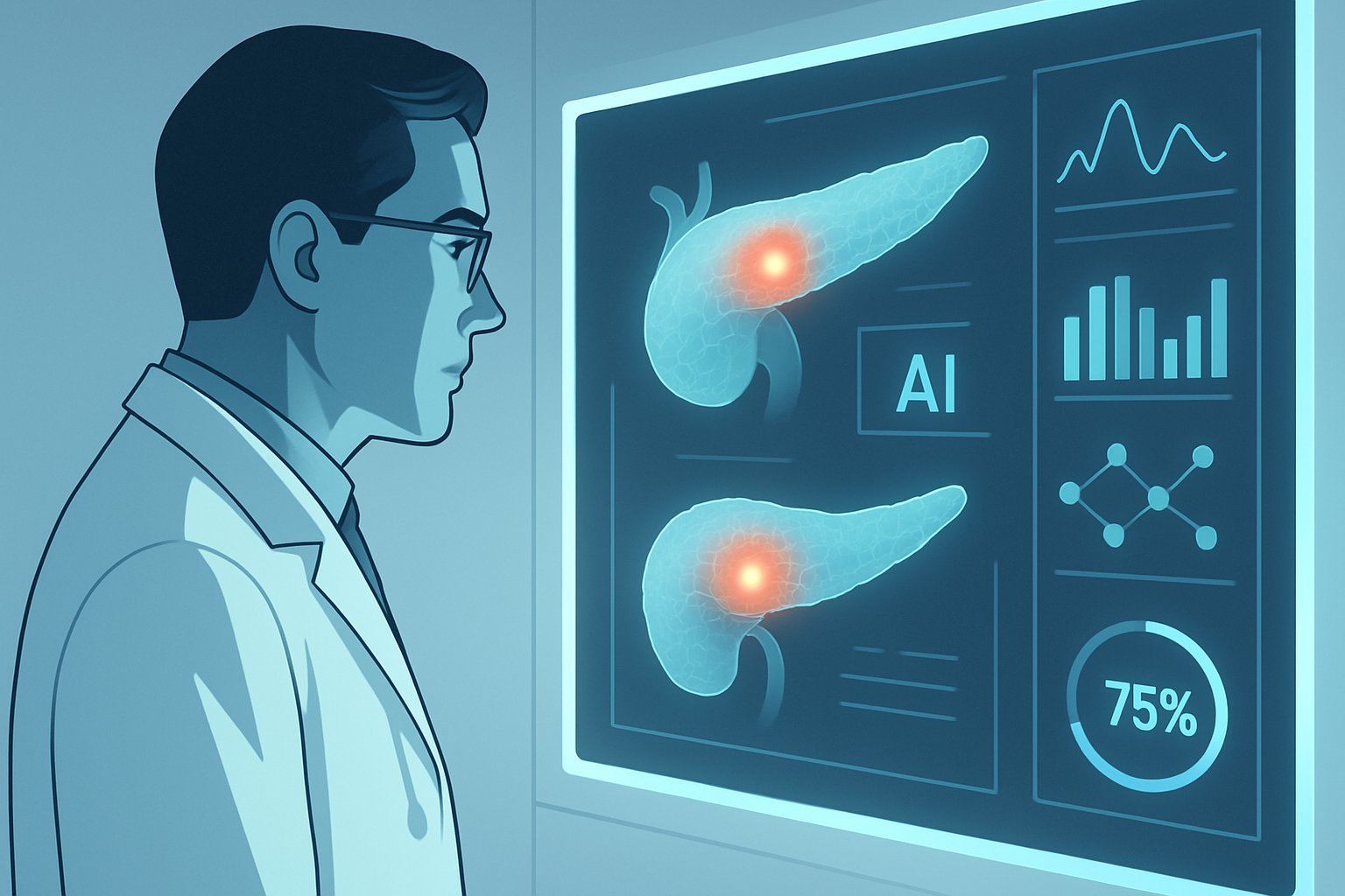
Taiwanese investigators soon replicated success using deep learning across a nationwide cohort of 1,473 CT examinations. Additionally, sensitivity remained near 90% while specificity approached 93% for tumors under two centimeters. Consequently, professional societies began discussing opportunistic Cancer Detection during unrelated abdominal imaging. Meanwhile, the Medical Imaging community is reevaluating routine CT workflows as potential screening assets. These data points fuel optimistic headlines, yet they also invite critical scrutiny.
Collectively, the studies suggest AI can surface occult disease far earlier than traditional review. However, deeper technical understanding clarifies how such performance is achieved.
Key Study Highlights Summarized
Three investigations dominate current discourse and numbers. Table-ready highlights help professionals compare them quickly.
- Gastroenterology 2022 radiomics: 95.5% sensitivity, 90.3% specificity, 0.98 AUC on test set.
- Radiology 2022 deep learning: 89.9% sensitivity, 95.9% specificity, nationwide validation AUC 0.95.
- Investigative Radiology 2025 PANCANAI: 91.8% sensitivity at diagnosis, 68.7% sensitivity months earlier.
Such Medical Imaging evidence tables help clinicians contextualize performance quickly. Importantly, only the Taiwanese study included thousands of controls, providing a realistic specificity estimate. In contrast, PANCANAI lacked non-cancer scans, preventing full accuracy calculations. Therefore, the 96% headline often references a single specificity value from internal or external subsets.
These distinctions emphasize that metrics vary by cohort, threshold, and prevalence. Next, we explore the algorithms driving those numbers.
Technology Behind This Breakthrough
Radiomics models extract hundreds of quantitative features describing organ texture, shape, and intensity. Subsequently, machine learning classifiers such as SVMs rank patterns that differentiate malignant from healthy tissue. Deep learning approaches instead feed pixel data into convolutional neural networks that learn hierarchical abstractions automatically.
Both strategies rely on precise pancreas segmentation, a historically difficult task due to organ variability. However, modern U-Nets deliver near-human delineation speed, enabling scalable Medical Imaging pipelines. Furthermore, many teams train on public datasets and then fine-tune using local scans for domain adaptation.
Hardware requirements remain modest; inference can run on existing Radiology workstation GPUs. Nevertheless, hospitals need robust data governance and cybersecurity safeguards for protected images.
Understanding model architecture helps stakeholders judge reproducibility and update pathways. The next section balances promise with practical clinical realities.
Benefits And Clinical Realities
Earlier Cancer Detection offers the chance for curative surgery before metastasis. Moreover, AI flags can guide enhanced surveillance for high-risk groups like familial history carriers. Automation also reduces radiologist fatigue during after-hours emergency interpretations.
Yet, false positives may trigger invasive endoscopic ultrasounds, anxiety, and added cost. Consequently, specificity must remain exceptionally high in low-prevalence screening populations. Prospective trials will define acceptable trade-offs and downstream care pathways.
Radiologists in busy Radiology suites must trust algorithms and understand operational boundaries.
Opportunities are clear, yet challenges can derail momentum without careful planning. Implementation considerations therefore loom large for health systems.
Implementation Roadmap For Hospitals
Leaders should begin with a structured pilot across multiple scanner vendors and contrast protocols. Additionally, they must prospectively collect outcomes to recalibrate thresholds as prevalence shifts. Below is a concise checklist for readiness. Embedding Medical Imaging analytics into PACS demands close vendor collaboration.
- Secure multidisciplinary governance including Radiology, surgery, oncology, and informatics.
- Integrate the algorithm within existing Medical Imaging PACS for seamless workflow.
- Define alert triage rules and follow-up timelines to limit unnecessary interventions.
- Monitor real-time performance dashboards and report drift to vendors promptly.
- Upskill staff through the AI in Healthcare™ certification.
Furthermore, health IT teams must test cybersecurity hardening before external data exchange. Meanwhile, payers will seek economic analyses demonstrating avoided late-stage treatment costs.
Thoughtful rollouts mitigate safety risks and build institutional confidence. Regulatory clarity will further accelerate adoption.
Medical Imaging Future Regulation
The FDA treats AI algorithms as Software as a Medical Device requiring clearance before marketing. Consequently, developers submit performance evidence and a Predetermined Change Control Plan outlining future updates. Updated guidance stresses transparency of Medical Imaging training data and post-market monitoring.
Europe follows the AI Act and existing MDR classifications, which demand risk management and human oversight. Meanwhile, standards groups are drafting benchmarking datasets to support fair Cancer Detection assessments. Global Medical Imaging societies are drafting consensus guidelines for transparent reporting. Hospitals must verify vendor claims with local validation to meet safety obligations.
Industry observers expect initial clearances focused on opportunistic rather than population screening use cases. Nevertheless, proactive certification and lifecycle planning will shorten review timelines.
Navigating global regulation requires technical documentation and clinical evidence. The final section synthesizes practical takeaways and future moves.
Conclusion And Next Moves
AI promises transformative, earlier pancreatic diagnosis through ever-improving Medical Imaging algorithms. Key studies demonstrate near 96% specificity and impressive sensitivity across diverse cohorts. However, most evidence remains retrospective, and real-world impact awaits prospective trials and regulatory clearance.
Healthcare leaders should launch controlled pilots, track performance, and prepare workforce reskilling. Therefore, the AI in Healthcare™ certification offers structured education for frontline teams. Consequently, institutions can translate cutting-edge research into measurable survival gains. Act now to pilot responsible AI, validate locally, and deliver hope to patients earlier.


